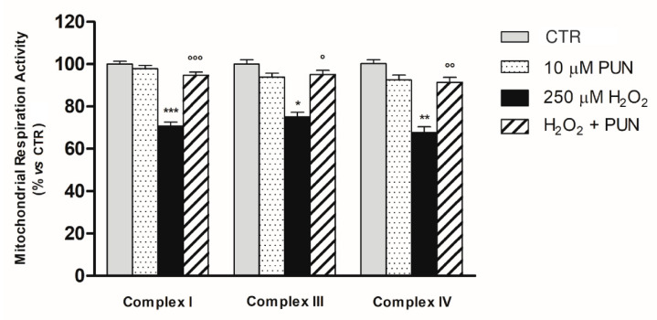
Figure 4.
The activities of mitochondrial respiratory chain complexes I, III, and IV were measured in mitochondria isolated of ARPE–19 cells. Enzyme activities were determined using specific assay kits (see Section 2.6 in Material and Methods). Results are shown as percentages relative to control for the single complex, which was set at 100%. Data are presented as the mean ± SEM of six replicates per experimental group from two independent experiments. One–way ANOVA analysis was carried out followed by post hoc Newman–Keuls test for each single respiratory complex. *, p < 0.05; ** = p < 0.01 and *** = p < 0.001 vs. Control; ° = p < 0.05; °° = p < 0.01 and °°° = p < 0.001 vs. H2O2 (250 µM) alone.

