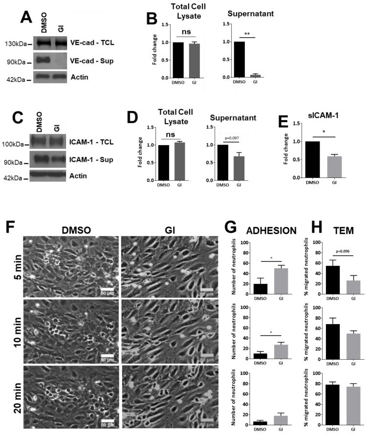
Figure 1.
ADAM10-regulated ICAM-1 Protein expression levels in HUVECs. (A) HUVECs cultured and treated as indicated (10 ng/mL TNFα) and analyzed on the expression of VE-cadherin by Western blotting. GI: ADAM10 inhibitor. Upper panel shows total cell lysate (TCL), middle one extracellular domain of VE-cadherin present in the supernatant of cultured ECs and lower panel shows actin as protein loading control for TCL. (B) Quantification of VE-cadherin expression in TCL and the extracellular domain of VE-cadherin present in the supernatant. (C) HUVECs were treated with inhibitors as indicated and ICAM-1 levels were analyzed in total cell lysates and endothelial cell supernatant by Western blotting, as described in A. Note that several ICAM-1 isoforms can exist ([26]. However, we did not find additional bands. (D) Same as under B but then for ICAM-1, TCL on left and extracellular ICAM-1 on the right. (E) Quantification of soluble ICAM-1 fragment (sICAM-1) in the supernatant measured by ELISA. (F) HUVECs were cultured in flow chambers and treated as indicated, followed by perfusion with neutrophils for indicated time points. Bar, 50 µm. (G) Quantification of number of neutrophils that adhered to TNFα-treated ECs, displayed for different time points as indicated under F. (H) Quantification of number of neutrophils that crossed TNFα-treated ECs (TEM: transendothelial migration), displayed for different time points as indicated under F. Data are mean of at least three independent experiments. * p < 0.05; ** p < 0.01.

