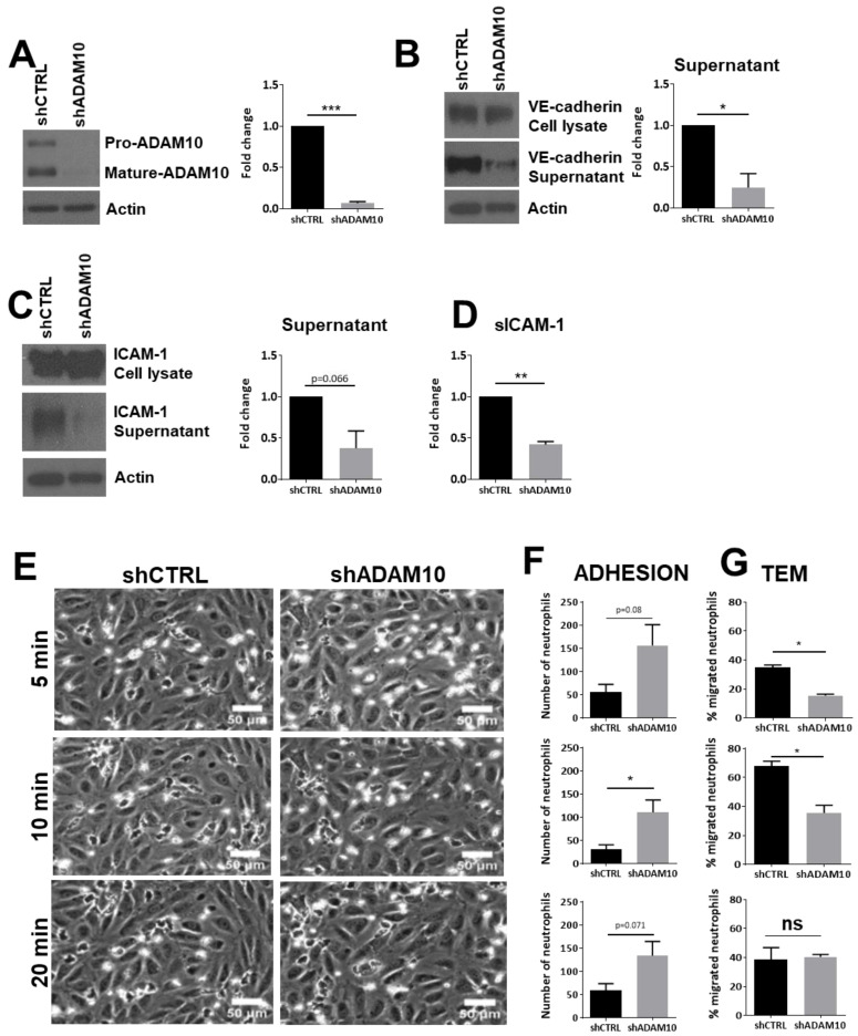
Figure 2.
Leukocyte transendothelial migration upon ADAM10 inhibition. (A) Cultured HUVECs were treated with shCTRL or shADAM10, checked by Western blotting. Actin was used as loading control. Quantification shows significant reduction of mature ADAM10 in ECs upon shRNA treatment. (B) ADAM10-deficient ECs show reduced shedding of VE-cadherin extracellular domain. Western blotting shows VE-cadherin expression in total cell lysate (upper panel), VE-cadherin extracellular domain in supernatant (middle panel) and actin as a loading control in lower panel. Quantification on the right shows significant reduction of extracellular VE-cadherin domain. (C) ADAM10-deficient ECs show reduced shedding of ICAM-1 extracellular domain, analyzed by Western blotting. Quantification on the right. (D) Soluble ICAM-1 levels were measured using ELISA. Reduced sICAM-1 in supernatant is measured in ADAM10-deficient ECs. (E) HUVECs were cultured in flow chambers and treated as indicated, followed by perfusion with neutrophils for indicated time points. Bar, 50 µm. (F) Quantification of number of neutrophils that adhered to TNFα-treated ECs, displayed for different time points as indicated under E. (G) Quantification of number of neutrophils that crossed TNFα-treated ECs, displayed for different time points as indicated under E. Data are mean of at least three independent experiments. * p < 0.05; ** p < 0.01; *** p < 0.001.

