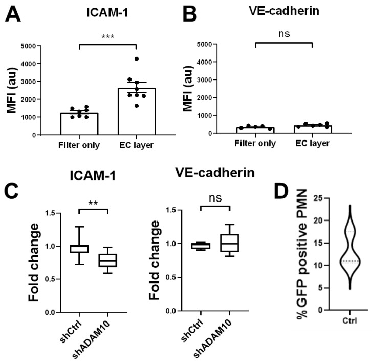
Figure 4.
Transmigration of neutrophil through endothelial monolayers. (A) Detection of ICAM-1 using antibodies that exclusively recognize extracellular part of ICAM-1 indicate that only neutrophils that crossed the endothelium (labeled as EC layer) show an increase of the presence of ICAM-1 on their surface, not when they crossed a bare filter (labeled as Filter only). (B) No increase in VE-cadherin is detected when using anti-VE-cadherin antibodies that detect the extracellular domain of VE-cadherin. (C) Silencing of EC-ADAM10 reduced the amount of ICAM-1 on the surface of the neutrophils compared to crossing shCTRL-treated EC monolayers, once they have crossed the endothelial monolayer. As a negative control, no change in VE-cadherin detection was measured (graph on right). Data are mean of at least three independent experiments. ** p < 0.01; *** p < 0.001. (D) HUVECs were transfected with ICAM-1-GFP, subsequently treated with TNF and neutrophils were allowed to cross ICAM-1-GFP-HUVECs in Transwell system. 10–15% of neutrophils that crossed transfected EC showed green signal, indicating membrane transfer during TEM. Experiment is carried out three times in duplicate.

