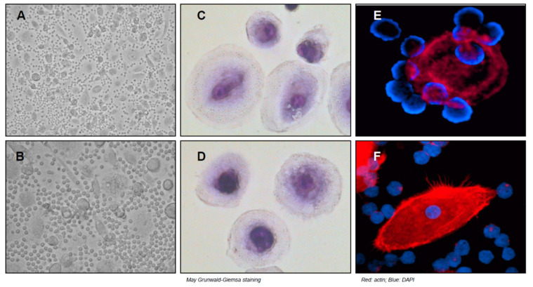
Figure 1.
NLC formation from the peripheral blood of patients affected by CLL. PBMC were isolated from CLL patients and cultured in complete medium for 14 days then CLL cells were carefully removed. (A–F) Figures with phase-contrast microphotographs, May–Grunwald–Giemsa, and immunofluorescence staining show the formation of large and adherent cells known as NLC. As shown, CLL cells are closely attached to NLC.

