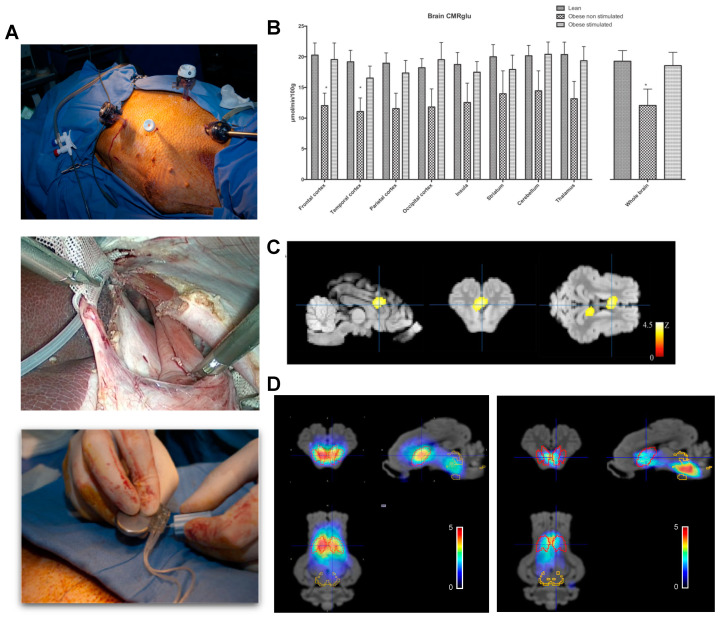
Figure 5.
Brain metabolism and SERT/DAT expression in adult miniature pigs after chronic abdominal vagal stimulation. (A). Laparoscopic access to the abdominal vagus at the level of the lower esophageal sphincter in the obese miniature pig. Two cuffs with electrode pairs Pt-Ir are located around each vagal nerve after careful dissection and subsequent closure of the esophageal groove. The electrode leads were connected to purpose made, double current high compliance channels, neurostimulator that was implanted in a subcutaneous pocket [109]. (B). Quantitative changes in brain glucose uptake after several weeks of chronic vagal stimulation in lean and obese animals showing restoration of obesity-related impaired glucose metabolism by VNS. (C). Voxel-based statistical parametric mapping analysis showing the differences in glucose metabolism between the obese non-stimulated and obese-stimulated groups. The image was centered the dorsal anterior cingular cortex, which was the region most markedly affected by stimulation. (D). Pixel-wise modeled SPECT dynamic image after administration of 123I ioflupane showing the binding potential of DAT/SERT overlaid on the MRI template. Red VOIs correspond to DAT-rich areas, whereas yellow VOIs represent SERT-rich areas. The left panel represents sham, whereas the right panel shows vagal stimulated obese miniature pigs. Adapted from Refs. [17,104].

