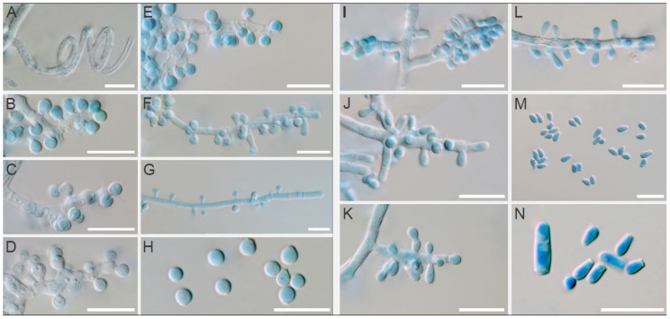
Figure 6.
Micromorphology of Trichophyton mentagrophytes (left; A–H) in comparison with T. erinacei (right; I–N) isolated from European hedgehogs in France. Spiral hyphae of T. mentagrophytes (A); branched or unbranched conidiophores bearing small round microconidia of T. mentagrophytes (B–G); free small round microconidia of T. mentagrophytes (H); simple conidiophores bearing microconidia of T. erinacei (I–L); free clavate microconidia and two-celled macroconidia of T. erinacei (M,N). Scale bars = 10 μm.

