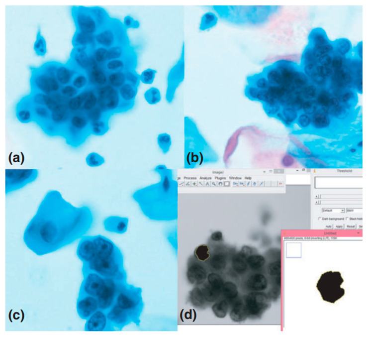
Figure 1.
The comparison of nucleus morphological features to identify cancer cells. Microphotographs (1000× magnification) of liquid-based cytology (LBC) cervical samples showing (a) atypical, (b) benign, and (c) malignant cells, and (d) screenshot from ImageJ software used to determine the selected morphometric parameters of cells [13]. Reproduced with permission from Gupta, P.; Gupta, N.; Dey, P., Cytopathology; published by John Wiley & Sons, 2017.

