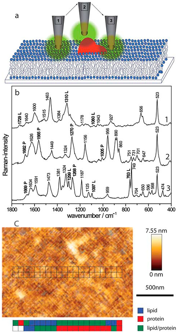
Figure 5.
The application of the tip-enhanced Raman spectroscopy (TERS) technique in the analysis of the chemical composition of biomembranes. (a) a representation of possible atomic force microscopy (AFM) tip positions corresponding to the measurement of interactions with lipid domains (1), protein domains (2) or both domains simultaneously (3); (b) TERS spectra corresponding with tip positions presented above. Bands characteristic of protein and lipid domains are marked with “P” or “L”, respectively; (c) AFM topography image of streptavidin-labelled phospholipid film with marked positions of particular domains detected with TERS [52]. Reproduced with permission from Böhme, R.; Cialla, D.; Richter, M.; Rösch, P.; Popp, J.; Deckert, V., J. Biophotonics; published by John Wiley & Sons, 2010.

