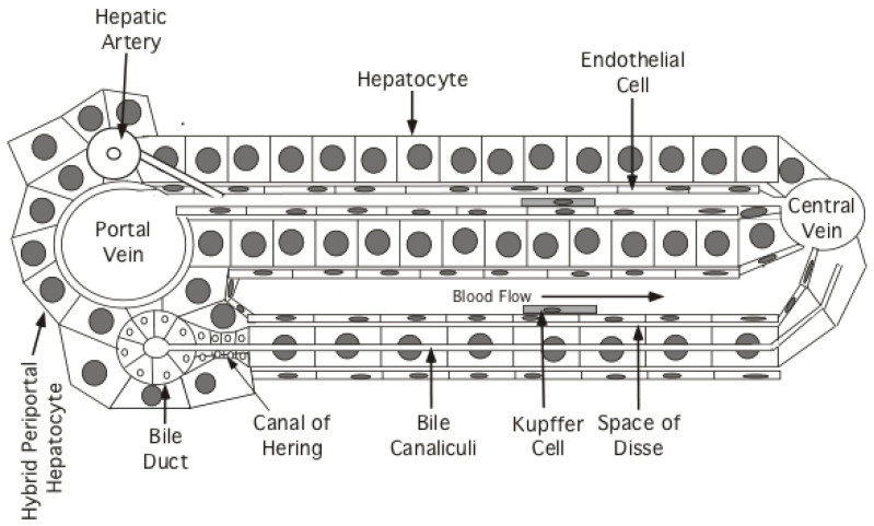
Figure 2.
Structure of the hepatic lobule: schematic, essentially one dimensional view, of the hepatic lobule, illustrating a few tracts of hepatocytes and supporting cells extending from a portal vein (in the portal triad) to a central vein. Hepatocytes or a subset of the hepatocytes constituting the limiting membrane around the portal triads (hybrid periportal hepatocytes), have the proliferative capacity to respond to severe liver injury and restore the hepatocyte population of the lobule [66]. Arterial and venous blood enters the sinusoids of the lobule at a portal triad and is collected at a central vein. Bile is secreted from hepatocytes into bile canaliculi, formed at the junction of adjacent hepatocytes, and flows into the bile ducts. The sinusoids are lined with a fenestrated endothelium; whether the fenestrations are large enough to allow free passage of HBV from the blood to hepatocytes is controversial, with an alternate suggestion that virus is actively transported from the blood stream to hepatocytes [71]. Kupffer cells (liver macrophages) are also shown, as well as the cells that form the Canals of Hering, which facilitate transport of bile from canaliculi to the bile ducts and have also been proposed to be hepatocyte progenitor cells [67]. Hepatic stellate cells (not shown), found within the Space of Disse, are multifunctional and, in the context of chronic HBV infection, are responsible for the development of cirrhosis in response to chronic liver injury. Figure adapted from Jilbert et al., 2008. Pathogenesis of HBV Infections, Chapter 7: HBV Human Virus Guide, 2nd Ed, CL Lai and S Locarnini Eds. Reproduced with permission from International Medical Press.

