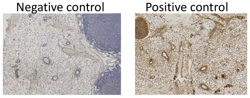
Figure A2.
Image showing examples of the negative and positive controls used in this study. The negative control was an antibody free control to determine any background staining caused by the kit used. The positive control was performed on tissue known to express the protein in question. These tests were performed on the same tissue types for each run performed, with this tissue being full adult, >16 weeks of age, wild type mammary glands.

