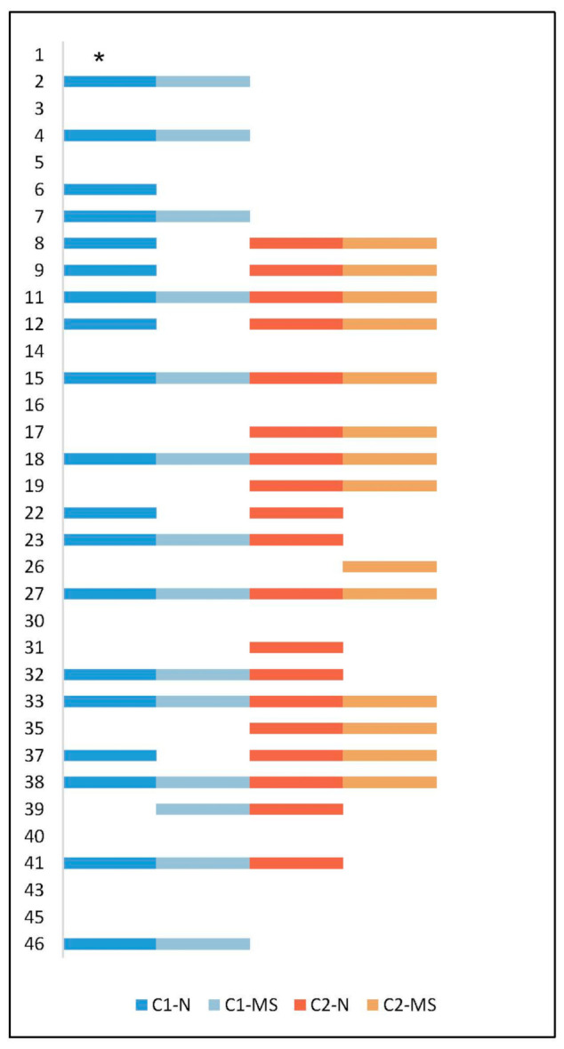
Figure 1.

Presence of Staphylococcus aureus in the nares and maxillary sinus in 34 patients at initial collection (C1) and at follow-up (C2) after approximately 11 years. The Y-axis shows the study ID for all 34 patients. One step on the X-axis symbolizes S. aureus isolated at a specific time point and site. Sample sites: maxillary sinus (MS), nares (N). * One sample from nares at C1 was not available.
