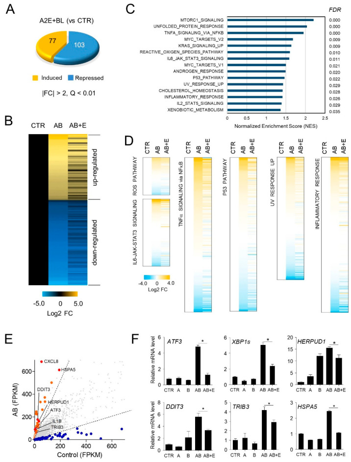
Figure 4.
Effect of EPX on genome-wide gene expression in ARPE-19 cells. (A) Differentially expressed genes by A2E and BL in ARPE-19 cells. A2E-laden ARPE-19 cells were exposed to blue light (BL), as shown in Figure 2A. RNA-seq analysis showed differentially expressed genes (DEGs) after BL exposure in A2E-laden RPE cells (A2E + BL) compared to untreated cells (CTR). FC—fold change; A2E—Bis-retinoid N-retinylidene-N-retinylethanolamine; BL—blue light. (B) Heatmap generated from three groups, including A2E-free ARPE-19 cells (CTR) and BL-stimulated A2E-laden RPE cells, in the absence (AB) and presence of EPX (AB + E). Values represent the log2 fold-change (FDR < 0.01) relative to CTR. (C–E) Pathways significantly altered after exposure to A2E and BL. From GSEA according to Hallmark collection, statistical significance in molecular functions (NES > 1, FDR < 0.05) is listed. AB—A2E+blue light; E—eggplant extract. (F) Validation of DEGs by RT-qPCR. ATF3—activating transcription factor 3; XBP1s—spliced X-box binding protein 1; TRIB3—tribbles pseudokinase 3; DDIT3—DNA damage-inducible transcript 3; HERPUD1—homocysteine-inducible endoplasmic reticulum protein with ubiquitin-like domain 1; HSPA5—heat shock protein family A (Hsp70) member 5. The results are presented as the mean ± S.D. (n = 4); * p < 0.05.

