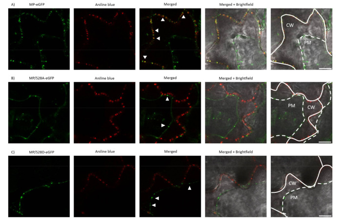
Figure 6.
Subcellular localization of wild type and mutant MP-eGFP constructs in aniline blue stained Nicotiana tabacum cv. Xanthi leaves. (A–C) Images were taken 24–48 h after agroinfiltration with a confocal microscope. For plasmolysis treatment, leaves were soaked in 10% NaCl solution immediately before examination. For better visualization, eGFP fluorescent and aniline blue images were false colored as green and red, respectively. The plasma membrane (PM) and the cell wall (CW) were marked by white dashed and continuous lines, respectively. Arrowheads indicate the punctate structures. Scale bar: 10 µm.

