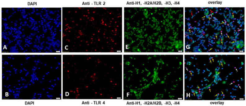
Figure 5.
Immunofluorescence analysis on bovine PMN activation of TLR2 and TLR 4 by E. bovis and concomitant neutrophil extracellular trap (NET) formation. PMN (n = 3; 5 × 10⁵) were exposed to vital E. bovis sporozoites (ratio 1:1) on poly-l-lysine-treated coverslips (120 min, 37 °C) and fixed for further antibody exposure (60 min) with anti-TLR2 (C) and anti-TLR4 (D) antibodies and anti-histone H1, H2A/H2B, H3, H4 antibody (E,F). Coverslips were mounted with ProLong Antifade containing DAPI (A,B) which was used for observation of PMN nuclei and NET extracellular DNA by fluorescence microscopy analysis. In both cases, expression of TLR2 and TLR4 was observed on the surface of bovine PMN (red) co-localized with NET-derived histones (green) and extracellular DNA (blue), as indicated by yellow arrows ((G,H)—overlay of images collected for nucleic acid, TLR2/TLR and histone staining). Co-localization of TLR-positive signals with early stages of NETosis are indicated by white arrows (G,H). TLR-positive signals without NETosis are indicated by orange arrows (G,H). Images were visualized by using an inverted Olympus IX81® epifluorescence microscope equipped with a digital camera (XM10®, Olympus, Tokyo, Japan). Scale bar magnitude: 20 µm.

