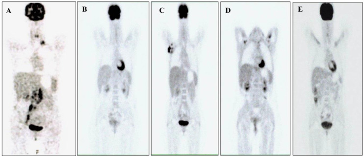
Figure 1.
(A) 18 F-fluorodeoxyglucose- Positron emission tomography–computed tomography ([18f] FDG-PET-CT) scans performed in October 2016; the examination showed pathologies with a high glucose metabolism in correspondence with multiple lymph node packets in the left supraclavicular region, retroclaveare left and right, left retrocrural, lomboaortica, intercavale, paracavale, the iliac bifurcation, and along the path of the internal inguinal vessels and right exteriors. (B) [18f] FDG-PET-CT scans performed in January 2018; the examination did not show pathologies with a high glucose metabolism. (C) [18f] FDG-PET-CT scans performed in July 2019; the examination showed pathologies with a high glucose metabolism in correspondence with numerous lymphadenomegalies in the right axillary region and of the lymph nodes in the right lung perilary area. (D) [18f] FDG-PET-CT scans performed in June 2020; the examination did not show pathologies with a high glucose metabolism. (E) [18f] FDG-PET-CT scans performed in September 2020; the examination did not show pathologies with a high glucose metabolism.

