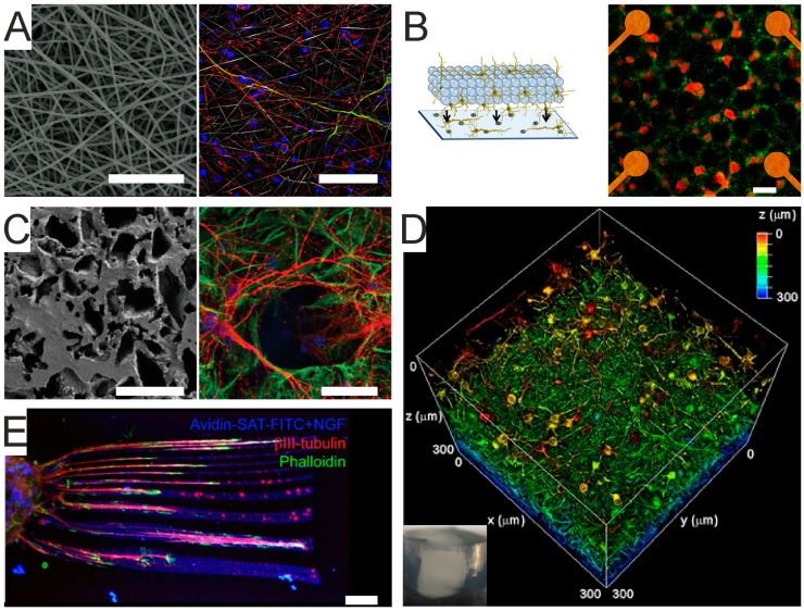
Figure 3.
Scaffold-based 3D cultures. (A) Electrospun fibers scaffold. Left—Scanning electron microscopy of thick electrospun fibers generated from tyrosine-derived polycarbonates. Scale bar 100 mm. Right—reprogramming induced pluripotent stem cells (iPSCs) on 3D electrospun fibers, leading to the generation of bIII-tubulin+ (red) and MAP2+ (green) neurons. Scale bar: 50 mm. Adapted from [79]. (B) Microbeads scaffold. Left—multilayered assembly of microbeads and primary neurons coupled with 2D primary neuronal cultures grown on a microelectrode array (MEA). Right - immunostaining of 3D culture on MEA, showing MAP-2+ (green) and NeuN+ (red) neurons. Scale bar: 40 μm. Adapted from [83]. Copyright © 2014 The Authors. (C) Graphene scaffold. Left—scanning electron microscopy image of a nanostructured PDMS–graphene scaffold. Scale bar: 200 m. Right—primary hippocampal neurons at 10 day-culture within the scaffold (green, betaIII tubulin+ neurons; red, GFAP+ glial cells). Scale bar: 50 μm. Adapted from [80]. Copyright © 2020 The Authors. (D) Alginate hydrogel scaffold. 3D reconstruction of a 300-μm3 volume of a cortical culture at 53 days in vitro. Color bar indicates the color-coded depth. The inset shows a macroscopic view of the bulk homogeneous alginate hydrogel. Adapted from [97]. (E) Hyaluronic acid hydrogel scaffold in which chick dorsal root ganglia axons are elongating within two-photon patterned microchannels functionalized with nerve growth factor. The bio-functionalized microchannels enable axon guidance within the hydrogel. Scale bar: 50 m. Adapted with permission from [86].

