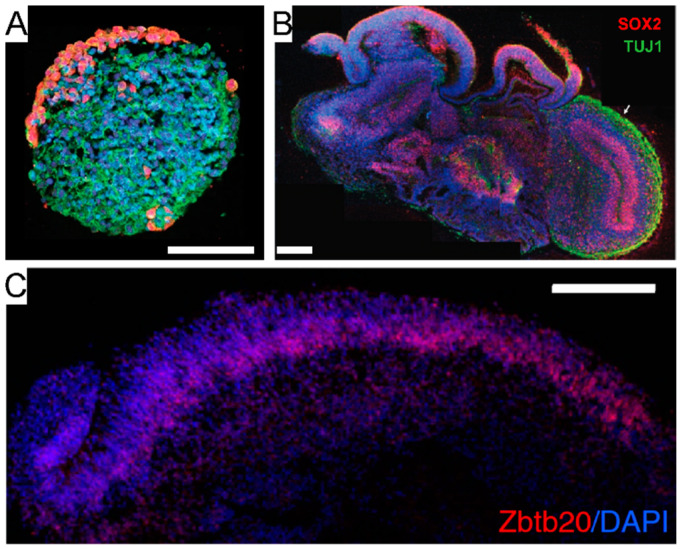
Figure 4.
Spheroids and organoids. (A) Brain spheroid. Human iPSC-derived brain spheroid using primary glioblastoma cells, stained for glia (GFP, red) and neurons (Tuj1, green). Scale bar: 100 μm. Adapted with permission from [121]. Copyright © 2019, The Authors. (B) Whole-brain organoid. Sectioning and immunohistochemistry reveal a complex morphology made of heterogeneous regions, and the presence of neural progenitors (SOX2) and neurons (TUJ1, arrow). Scale bar: 200 μm. Adapted with permission from [124]. Copyright © 2013, Nature Publishing Group. (C) Region-specific organoid. Hippocampus-like tissue expressing the specific marker Zbtb2. DAPI: nuclei. Scale bar: 100 μm. Adapted with permission from [133]. Copyright © 2015, The Authors.

