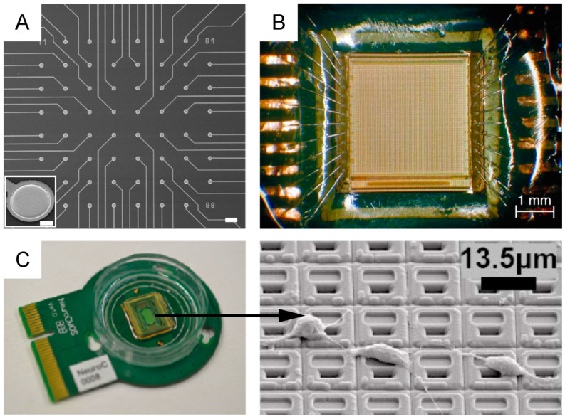
Figure 8.
Active and passive planar MEAs. (A) Passive planar MEA (scale bar: 100 μm). The inset shows the scanning electron microscopy image of a TiN microelectrode (scale bar: 10 μm. Adapted with permission from [203]. Copyright © 1998 Elsevier Science B.V. (B) Active pixel sensor MEA made of 4096 gold microelectrodes. From [246] with permission. Copyright © 2004 Elsevier B.V. (C) High-density (HD)-MEA with ~60000 electrodes. Left—biocompatible chip packaging and PCB. Right—scanning electron microscopy image of the chip surface, showing in-house post-processed Pt-electrodes and dissociated primary rat cortical neurons, cultured on top. Adapted with permission from [249].

