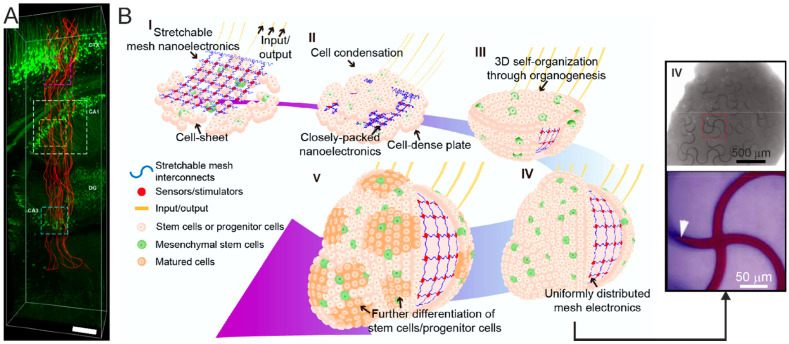
Figure 14.
Mesh electronics for tissue-wide electrophysiology. (A) Injectable mesh electronics. 3D reconstruction of the interface between neurons (green) and the injected electrodes mesh (red) at 6 weeks post-implantation. The mesh spans the cortex and reaches the hippocampus. Scale bar, 200 μm. CA1: Cornu Ammonis 1. CA3: Cornu Ammonis 3. DG: Dentate Gyrus. Adapted with permission from [291]. (B) Left—schematic rendition of the stepwise assembly (I-IV) of the mesh electronics into the organoid through organogenesis. Right—bright-field image of a fully assembled organoid incorporating unfolded mesh electronics (step IV in the schematic rendition) and zoomed-in view of the region marked by the dashed red square. Adapted with permission from [143]. Copyright © 2019 American Chemical Society.

