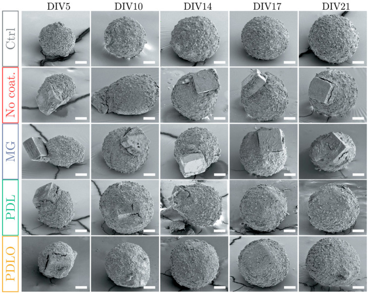
Figure 15.
Functionalization-driven self-standing micro-device integration in 3D neural cultures. Scanning electron microscopy images of fixed neurospheroids taken at different days in vitro (DIV), showing the incorporation time course of functionalized and non-functionalized self-standing micro-devices. Ctrl: control condition, neurospheroid without microchip. No coating: non-functionalized micro-device. MG: matrigel. PDL: Poly-D-lysine. PDLO: poly-DL-ornithine. When the microchip is either non-coated or coated with MG, it remains at the periphery of the spheroid, whereas it is progressively incorporated within the spheroid when coated with either PDL or PDLO. Scale bar: 50 µm. From [302].

