Figure 1.
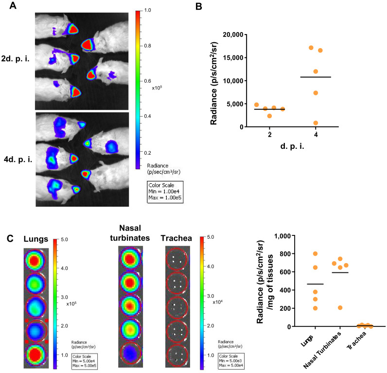
RSV replication in whole BALB/c mice. Adult BALB/c mice were intranasally infected with rHRSV-Luc. (A) At 2- and 4-days post-infection (d.p.i.), mice intranasally received luciferin (1.5 mg under 50 µL), and luciferase activity was visualized in the IVIS-200 imaging system. Images make it possible to observe virus replication in the nose and the lungs of animals at 2- and 4- days post-infection. The scale on the right indicates the average radiance: the sum of the photons per second from each pixel inside the region of interest/number of pixels (p/s/cm2/sr). A red signal indicates a high radiance, and consequently a high luciferase activity, whereas a blue signal indicates a low radiance, indicative of a low replication. (B) Quantification of luciferase activity in the lungs of living mice (radiance in photon/s/cm2/sr). (C) Quantification of luciferase activity in lungs, nasal turbinates (NT) or trachea homogenates. The scales indicate the average radiance: the sum of the photons per second from each pixel inside the region of interest/number of pixels (p/s/cm2/sr). Individual results were expressed radiance in photon/s/cm2/sr/mg of tissues and means are expressed as mean of individual ± SEM (one representative experiment of two with n= 5 to 8 mice/group) are represented (n= 5 mice/group).

