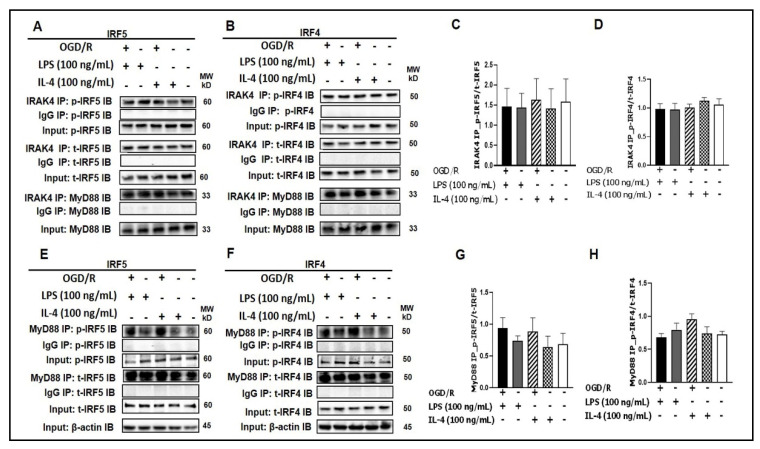
Figure 1.
IRF5 and IRF4 bind to MyD88 or IRAK4. (A, B) SIM-A9 cell homogenates were subjected to Co-IP with anti-IRAK4 antibody followed by immunoblotting for p-IRF5/p-IRF4, t-IRF5/t-IRF4, and MyD88. (C, D) WB optical density quantification of the ratio of p-IRF5 over t-IRF5 (C) and p-IRF4 over t-IRF4 (D). (E, F) SIM-A9 cell homogenates were subjected to Co-IP with anti-MyD88 antibody to detect p-IRF5/p-IRF4 and t-IRF5/t-IRF4. (G, H) WB optical density quantification of the ratio of p-IRF5 over t-IRF5 (G) and p-IRF4 over t-IRF4 (H). IgG controls were from the same homogenates of each treatment. n = 3 independent experiments/per condition. IB, immunoblot; p, phosphorylated; t, total; IgG, immunoglobulin negative control. One-way ANOVA.

