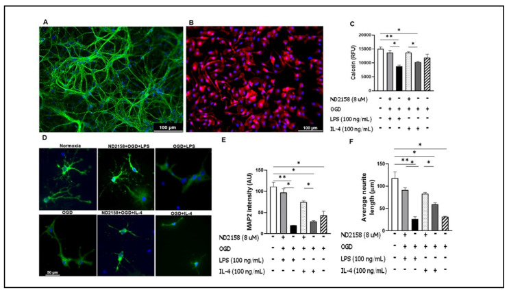
Figure 5.
Morphological changes and viability of neurons treated with conditioned medium of microglial culture. Cell culture media from primary microglia treated with ND2158 and stimulated with OGD + LPS or +IL-4, were used to re-perfuse neuronal culture of E16–18 for 24 h. (A) Mature neurons stained with DAPI and MAP2 antibody. (B) Cultured microglia stained with DAPI and Iba-1. Neuronal morphological changes after exposure of neurons to the conditioned media, and quantification of neurite lengths and MAP2 intensity. (C) Neuronal viability quantification with Calcein assay. (D) Neuronal morphology after exposure to conditioned microglia culture medium. Quantification of MAP2 fluorescence intensity (E) and neurite lengths (F) in (D). Data were averaged from total 9–12 images in three independent experiments; * p < 0.05; One-way ANOVA (D); Scale bar = 100 µm (A,B)/50 µm (D).

