Figure 1.
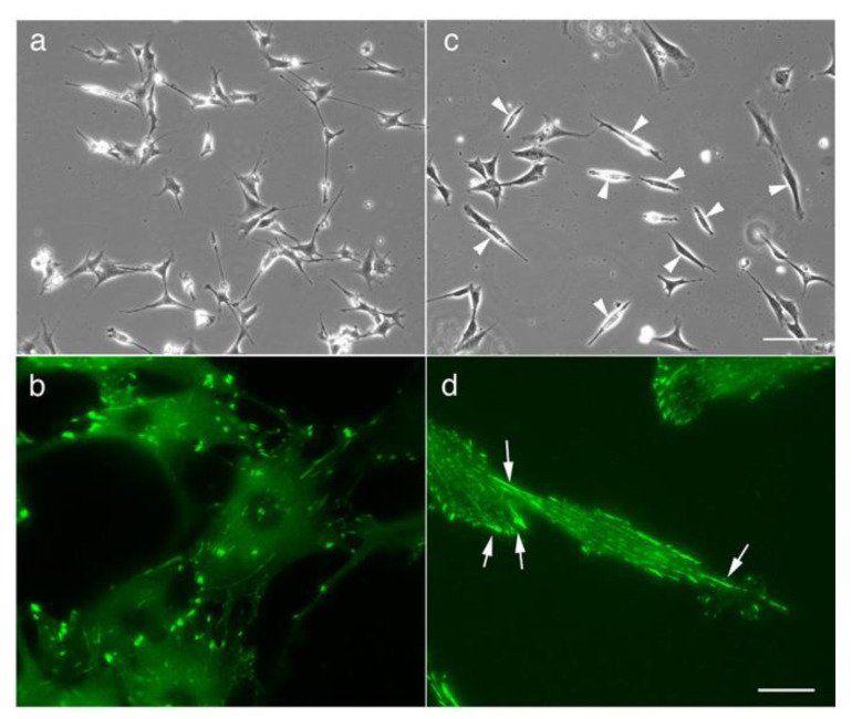
Morphology of normal 3T3 cells and Src knockout fibroblast (SYF) cells (c-Src, c-Yes, and Fyn knockout cells), as observed by phase-contrast microscopy. (a) The morphology of normal fibroblasts. (b) Fluorescence microscopy showing focal adhesions stained with anti-vinculin antibody. (c) When SYF cells were cultured on a glass substrate, they first showed a pancake-like morphology and then adopted a symmetrical spindle shape (arrowheads). (d) In this process, focal adhesions were formed at both ends of the cells, and a relatively small adhesive patch-like structure was observed at the center of the cells (arrows). (a,c) Phase-contrast microscopy. (b,d) Fluorescence microscopy showing focal adhesions stained with anti-vinculin antibody. Scale bars: (a,c), 100 μm; (b,d), 20 μm.

