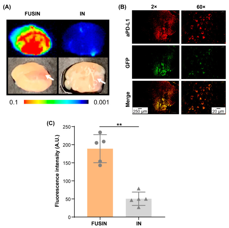
Figure 4.
FUSIN delivery of 800CW-aPD-L1 to the brainstem glioma. (A) Fluorescence images of representative ex vivo mouse brainstem slices. The white arrow indicates the tumor location. (B) Spatial distribution of FUSIN-delivered 800CW-aPD-L1. Left panel: aPD-L1 distribution in a coronal section of the brainstem after FUSIN delivery imaged at 2×. Right panel: higher magnification view (60×) of the tumor showing the colocalization of aPD-L1 with the tumor cells. (C) Fluorescence quantification of the 800CW-aPD-L1 delivery efficiency to the brainstem glioma (** p < 0.01).

