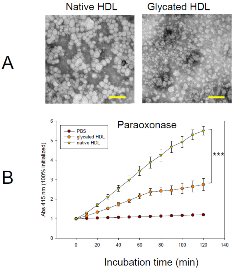
Figure 2.
Observation of the HDL particle and antioxidant activity. (A) Negatively stained image of HDL from transmitted electron microscopy at a magnification of ×40,000. Scale bar (yellow) indicates 100 nm. (B) Changes in the activity of paraoxonase in HDL with or without glycation. The error bars indicate the SD from three independent experiments with duplicate samples. The molar extinction coefficient of p-nitrophenol was 17,000 M−1 cm−1. ***, p < 0.001.

