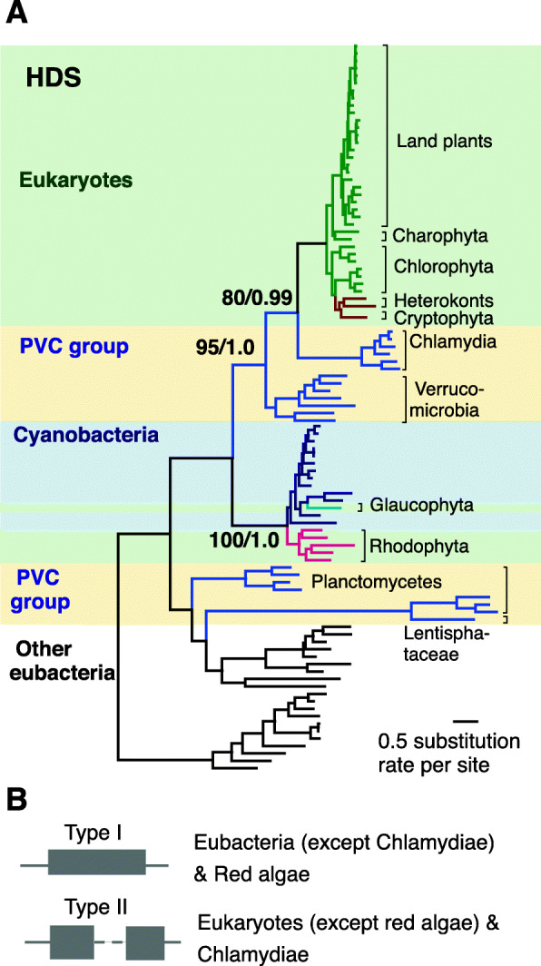
Fig. 3.

Origin and domain structure of HDS. a Cladograms cluster plastid-bearing eukaryotes HDS with chlamydia amino acid sequences. The cladograms are obtained from RAxML using amino acid sequences. Taxa in different major groups are shown in different colors. Species in plastid-bearing eukaryotes, PVC groups and Cyanobacteria are shaded with light green, light yellow and light blue, respectively. Numbers associated with branches are bootstrap (BS) values obtained from RAxML and posterior probability are obtained from MrBayes. b Schematics of the types (type I and type II) of gcpE domain in HDS enzyme and their presence in their corresponding species
