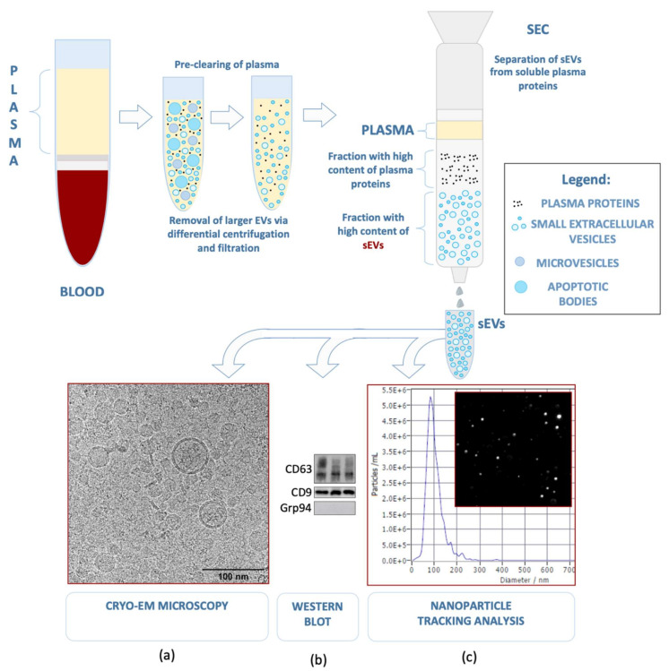
Figure 1.
Small extracellular vesicles present in human plasma may be separated using pre-clearing with differential centrifugation and a 200 nm filter (not shown), followed by size exclusion chromatography. The isolated sEVs can be characterized according to the guidelines of the International Society for Extracellular Vesicles [4] with the use of (a) Cryo-EM microscopy (52,000×) to estimate their size and morphology, (b) western blot for two positive (CD63 and CD9) and one negative (Grp94) sEV marker, and (c) nanoparticle tracking analysis (NTA), which allows the evaluation of vesicle size (average diameter = 90.9 nm) and concentration (1.3 × 10¹¹ particles/mL) [9] (modified).

