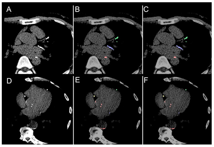
Figure 4.
Comparison. (A,D) Illustrative comparisons between original image; (B,E) manual annotation ground truth; (C,F) the model predicted segmentation mask. The model is able to identify both calcifications of the left anterior descending (green), left circumflex (blue) and right coronary arteries (yellow) as well as aortic calcium (red).

