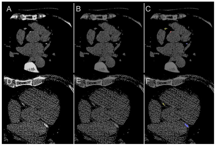
Figure 6.
Potential for Error Detection. Illustrative examples of the model as a ‘second reader’, correcting the human error. (A,D) The original images showing calcification of the left circumflex (blue) and right coronary arteries (yellow), as well as aorta (red); (B,E) manual annotation, showing missed calling of calcification; (C,F) correct identification of calcifications that were missed by manual annotation.

