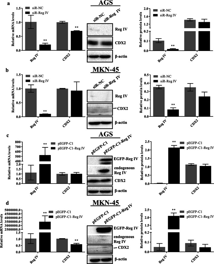
Fig. 4.
Reg IV had no effect on CDX2 in gastric cancer cells. AGS and MKN-45 cells were transfected with siR-Reg IV (a, b) or pEGFP-C1-Reg IV (c, d). Reg IV and CDX2 mRNA levels were evaluated using real-time PCR, and relative mRNA expression results were normalized to GAPDH (left of a–d). Reg IV and CDX2 protein expression levels were analyzed via western blotting. β-actin was used as a loading control. Representative immunoblots are shown in the middle panels of a–d. The relative protein signal intensity was quantitatively analyzed using Image Lab software and shown as histograms (right of a–d). Full-length original blots are presented in Additional file 1. Data are presented as mean ± SD. *p < 0.05; **p < 0.01 compared with the control groups

