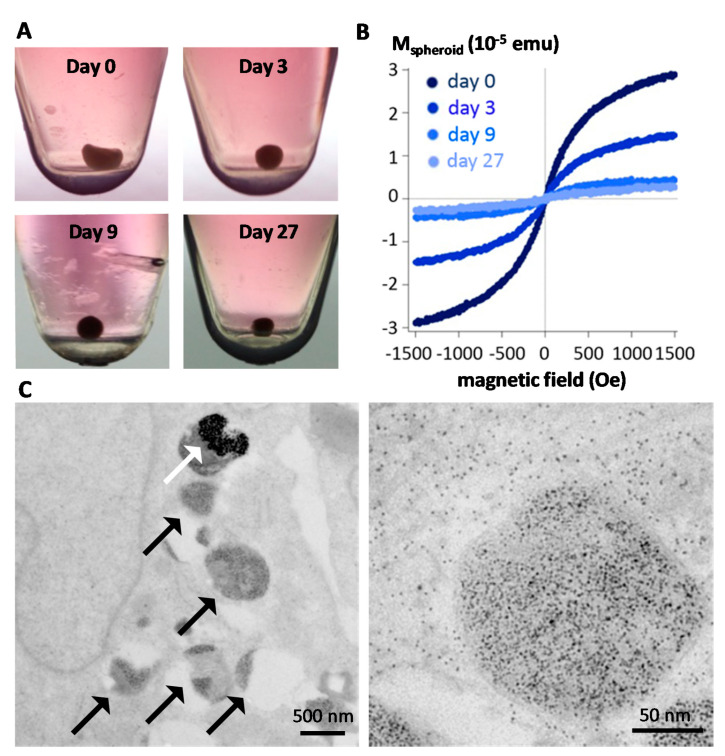
Figure 3.
Intracellular biodegradation of MNPs monitored within cell spheroids as tissue model. (A) The spheroids are formed from 200,000 stem cells, which spontaneously regroup into a cohesive spherical aggregate that can be kept in culture over a month without experiencing any cell mortality or tissue necrosis. (B) Magnetometry can be performed at the single spheroid level, evidencing a massive degradation of the nanoparticles in a few days after internalization. (C) TEM images one month after nanoparticle internalization, demonstrating that only a few intact nanoparticles remain within the endosomes (white arrow) while both endosomes and cytoplasm are filled with the ferritin protein (black arrows) containing the iron released from degradation, with a diameter 5–7 nm as seen in the close-up image on the right. Reproduced with permission from [111].

