Fig. 1.
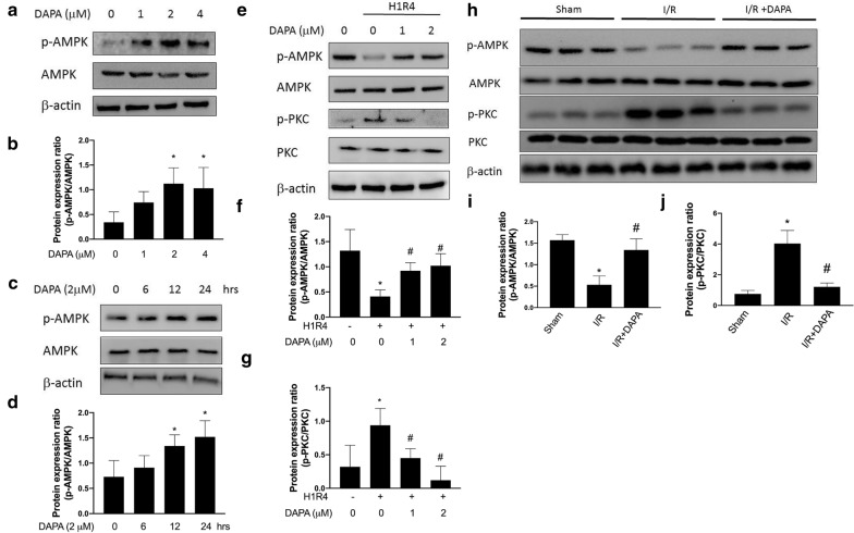
Administration of dapagliflozin (DAPA) increases the phosphorylation of AMPK and suppresses the phosphorylation of PKC under hypoxia/reoxygenation condition.
The expression levels of phosphorylated AMPK in H9c2 cells treated with DAPA were enhanced in dose-dependent (a, b) and time-dependent (c, d) manners. Representative Western blot images (e) and relative densitometric bar graphs of phosphorylated-AMPK/AMPK (f) and phosphorylated-PKC/PKC (g) in H9c2 cells exposed to hypoxia for 1 h and reoxygenation for 4 h (H1R4) were shown. The data were presented as the mean ± SD of three biological replicates at three separate times. Representative Western blot image (h) and protein expression levels of phosphorylated-AMPK, AMPK, phosphorylated-PKC, and PKC in ventricular tissue from sham control, ischemia/reperfusion (I/R), and I/R plus DAPA treatment animals, eight animals in each group, were shown (i, j). (* indicating p < 0.05 compared with the control group; # indicating p < 0.05 compared to H1R4 condition or I/R without DAPA treatment)

