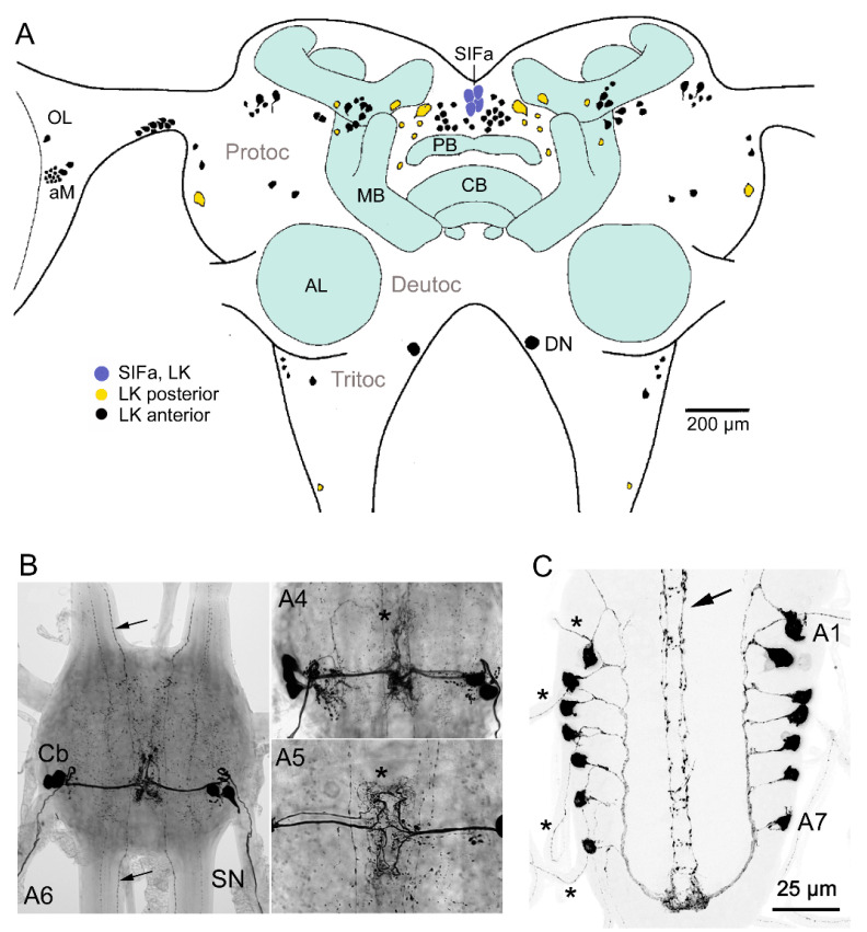
Figure 6.
LK neurons in the CNS of the locust. (A) LK immunoreactive neurons in the brain of the locust L. migratoria. LK cell bodies are predominantly found in protocerebrum (Protoc), including the optic lobes (OL) and accessory medulla (aMe; pacemaker region of clock), but some are in tritocerebrum (Tritoc). Neuronal process from LK neurons (not shown) are in the central body, optic lobe, and antennal lobe (AL), and less delineated neuropils are shown in all three brain neuromeres. A group of four neurons (SIFamide producing (SIFa)) in the pars intercerebralis coexpress SIFamide and LK. These SIFa neurons are known to send processes throughout the brain and ventral nerve cord [83,84] similar to the four SIFa neurons in Drosophila [85]. (B) LK immunoreactive neurons (neurosecretory cells) in abdominal ganglia of the locust Locusta migratoria. In more anterior abdominal ganglia (A1–A4), there are three pairs of LK neurons, and in the posterior ones (such as A6 shown here), there are only two pairs. The arrows depict axons from the descending neurons (a total of four axons). Other abbreviations: Cb, cell body; SN, segmental nerve. (C) In larval Drosophila, there is one pair of LK immunoreactive ABLKs (abdominal ganglion LK neurons) in each of the abdominal neuromeres A1–A7. These send axons to the body-wall muscle via segmental nerves (asterisks). Arrow indicates axons of the two pairs of descending neurons, SELK. Panel A is altered from [34] with SIF neurons added [83], B is from [86], and C is altered from [87]. All figures used with permission from publishers.

