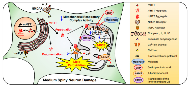
Figure 3.
Mitochondrial dysfunction and lipid peroxidation are found in the medium spiny neuron (MSN) of HD. mutant Huntingtin (mHTT) gene expresses mHTT protein in the MSN. mHTT inclusions are increased in the nucleus and cytosol. In the cytoplasm, both increased mHTT protein fragments and aggregates triggers neuronal damage. The aggregated form of mHTT decreases Krebs cycle and Ca2+ activity, resulting in a decrease in complex I–IV or mitochondrial respiratory complex activity. mHTT aggregation also decreases mitochondrial membrane potential (ΔΨ) and increases reactive oxygen species (ROS) production. As Ca2+ permeability increases, the mitochondrial membrane is hyperpolarized, triggering cell loss. In chemical models of Huntington’s disease (HD), malonate and 3-nitropropionic acid (3NP) inhibits succinate dehydrogenase (SDH) at complex II and, eventually, reduces ATP production via complex V. Lastly, the level of 4-hydroxynonenal (4-HNE), a neuronal lipid peroxidative damage marker, is elevated in HD and nordihydroguaiaretic acid (NDGA) reduces 4-HNE levels and prevents mitochondrial damage in the MSN.

