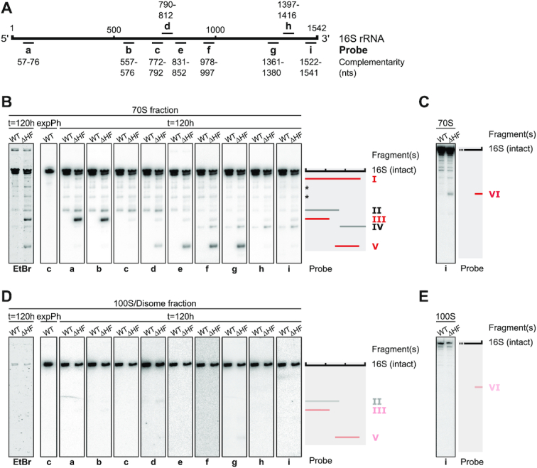
Figure 3.

ΔHF cells accumulate 70S particles containing fragmented 16S rRNA. (A) Schematic of 16S rRNA and DNA oligonucleotide probes used for Northern blot analysis in (B-E). For the sequences of the indicated probes see Supplementary Table S2. (B) Northern blot analysis of 16S rRNA fragments found in the 70S fraction of five day old WT and ΔHF cultures. For orientation a cut-out of the ethidium bromide-stained gel (Supplementary Figure S4A) used for Northern blotting is shown (left-most panel). RNA purified from the 70S fraction of an exponentially growing WT culture served as a control for the detection of intact 16S rRNA. The schematic on the right shows fragments (I–V) mapped by urea–PAGE and Northern blot analysis. Gray bars indicate fragments common to both WT and ΔHF-70S particles (fragments II and IV), red bars indicate fragments only present in ΔHF-70S particles (fragments I, III and V). Asterisks mark additional unidentified 16S fragments or unspecific binding of labelled oligomers to fragments of 23S rRNA. (C) As described in (B). RNA was resolved on 6% urea–PAGE gels for detection of RNA in a lower size range. (D) Northern blot analysis of 16S rRNA found in the 100S/disome fraction of five day old WT and ΔHF cultures, as described in (B). Traces of fragments II, III and V are indicated by opaque bars in the schematic. (E) As described in (D). RNA was resolved on 6% urea–PAGE gels for detection of RNA in a lower size range.
