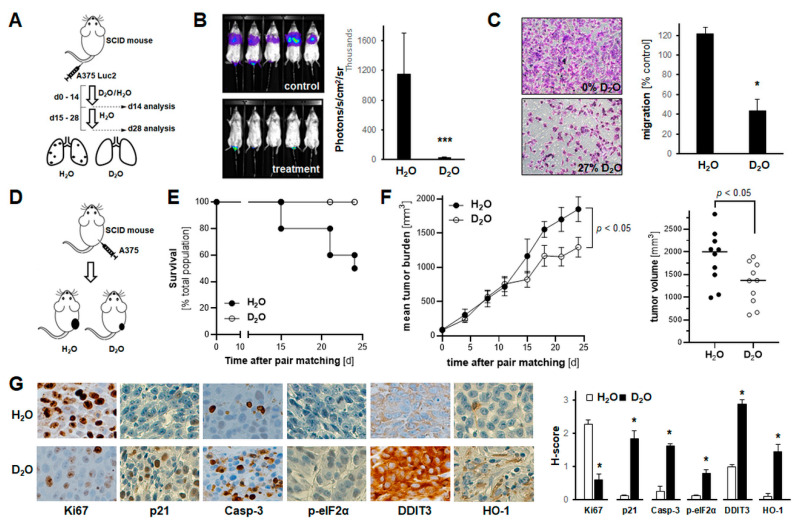
Figure 5.
Systemic administration of D2O attenuates A375 melanoma cell invasiveness in vitro, while impairing metastasis and tumor growth in SCID mouse models of human malignant melanoma. (A,B) A375-Luc2 melanoma cells were tail vein injected (n = 8 per group) followed by bioluminescent image analysis of lung metastasis (14 and 28 days later). Starting at time of cell injection until day 14, mice received H2O-based or D2O-supplemented (30% v/v in H2O) drinking water, followed by 14 days H2O in both groups. A, injection scheme; B, bioluminescent imaging (day 28) with bar graph depicting numeric image analysis of bioluminescent signal (p *** < 0.001). (For bioluminescent imaging on day 14, see Figure S4). (C) Invasion through Matrigel-coated Boyden chamber (H2O-based versus D2O-supplemented (27%) medium). Bar graphs with representative images (10× magnification) after crystal violet staining of inserts (n = 3; p * < 0.05). (D–G) A375 melanoma cells were injected subcutaneously (n = 10 per group); after pair-matching, tumor growth was monitored over a 24-day period; starting at time of pair matching (day 0) until end of experiment (day 24), mice received H2O-based or D2O-supplemented (30% v/v in H2O) drinking water. (D) Experimental scheme. (E) Kaplan–Meier analysis of mouse survival as a function of treatment groups; numbers indicate survivors per group. (F) Tumor burden in treatment groups as a function of time; graph (right panel), individual tumor size at termination. (G) At the end of the experiment, tumors were processed for IHC (left panels; 20× magnification); bar graph: tissue H-scores per antigen (right panel; n = 3; p * < 0.05).

