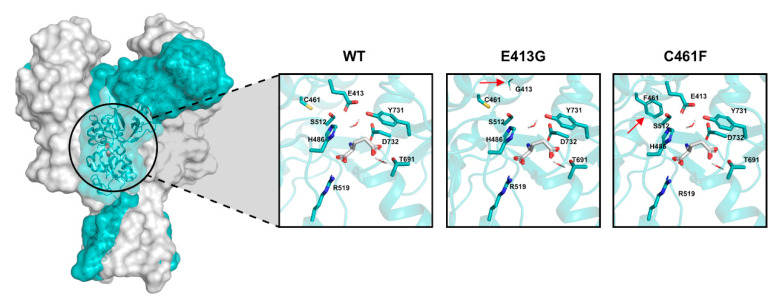
Figure 9.
Structure of human GluN1/GluN2A NMDA receptor (PDB accession code: 4TLM). The GluN2B subunit is colored in light blue. The insights show the glutamate binding domain of the wild-type (WT) receptor and the two structural variants Glu413Gly (E413G) and Cys461Phe (C461F). Each window focuses on the docked glutamate (white molecule) and the crucial residues that directly participate to the interaction. Red arrows point to the residue substitution of each of the two structural variants.

