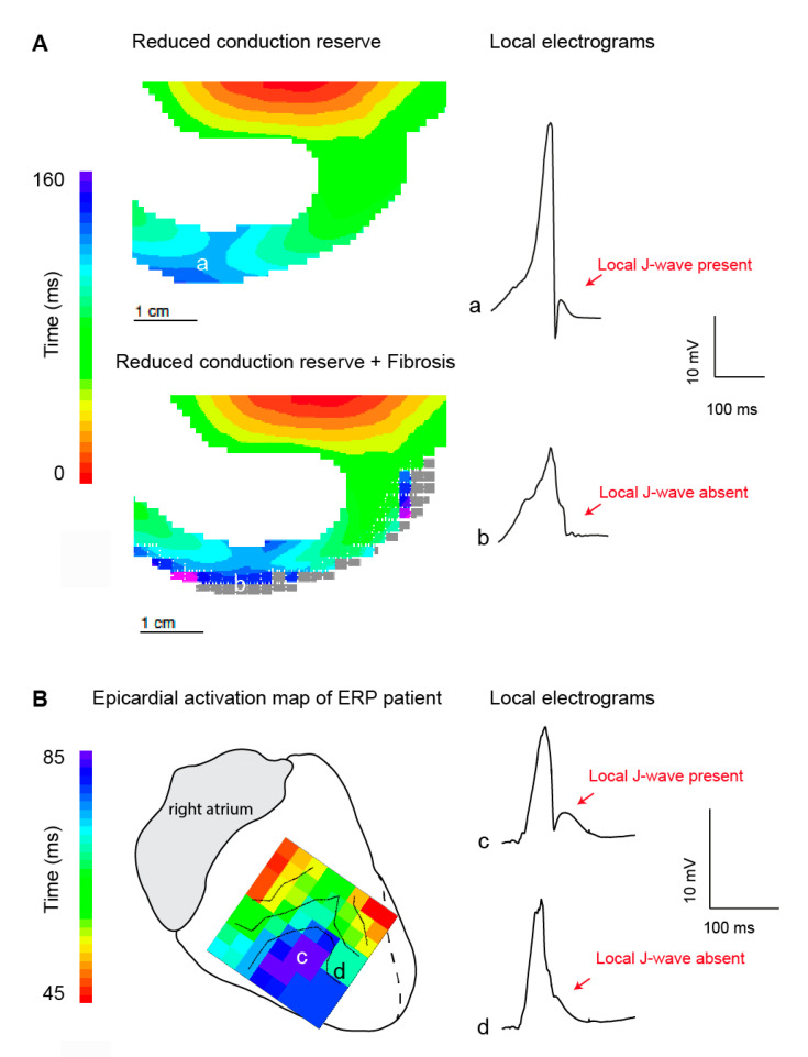
Figure 2.
Local J-wave and excitation failure in relation to the early repolarization syndrome. (A). Simulated activation maps and extracellular potentials during sodium channel reduction (30%) in the absence (upper) and presence (lower) of structural abnormalities. Propagation and extracellular potential were simulated using a similar approach as published previously [12]. In brief, propagating action potentials (AP) were simulated with a monodomain reaction-diffusion equation, using software that has been described previously [40]. Ionic currents were computed with the Ten Tusscher-Noble-Noble-Panfilov model for the human ventricular myocyte [41], which distinguishes subendocardial, mid-myocardial, and subepicardial cell types. Computation of extracellular potentials (electrograms) from the simulated membrane potentials was based on the bidomain model for cardiac tissue [42]. Delayed activation in location a resulted in a pronounced J-wave in the corresponding local electrograms. In the presence of structural abnormalities excitation failure occurred and the local J-wave disappeared. Panel (B) shows the epicardial activation pattern at the right inferior wall measured during open chest mapping [16]. The delayed activated myocardium (in blue) showed unipolar electrograms with (c) and without (d) a local J-wave. We speculate that excitation failure occurred at location d. ERP, early repolarization pattern.

