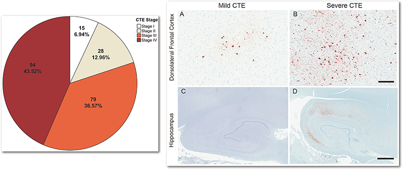
Fig. 5. Dementia Status by CTE Stage and p-tau Accumulation in the Dorsolateral Frontal Cortex and Hippocampus.
Left: Pie chart of the number (%) of brain donors with dementia by CTE stage (N = 359). For percents, denominator is total brain donors with dementia (n = 216). Right: In CTE, p-tau deposition often begins in the dorsolateral frontal cortex (DLF), with the hippocampus becoming involved in later disease stages (i.e., Stage III). The density of p-tau in the DLF and hippocampus and the size of the pathognomonic CTE lesions increases with age. These regions are therefore sensitive markers of the progression and severity of disease. The images on the right are exemplary of the severity of pathology in these regions by age and dementia status. (A) Mild perivascular accumulation of p-tau at the depths of the cortical sulcus in the DLF in a non-demented 30 year old with stage II CTE; (B) Severe perivascular accumulation of p-tau at the depths of the sulcus in the DLF in a demented 80 year old with stage IV CTE; (C) Absence of hippocampal p-tau accumulation in a non-demented 44 year old with stage II CTE; (D) Hippocampal p-tau accumulation in a demented 69 year old with stage III CTE. Positive staining for p-tau is depicted in red while hematoxylin counterstain is in blue. Scale bar represents 200um (A, B) and 2mm (C, D).

