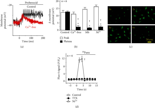
Figure 3.

T-type voltage-dependent Ca2+ channels (T-type Cav) are involved in the endothelial cell depolarization evoked by the Panx-1 channel blockade. (a) Representative recordings of the changes in endothelial cell membrane potential evoked by 1 mM probenecid in control conditions or after removing Ca2+ ions from the buffer solution (Ca2+-free solution plus 2 mM EGTA). Note that the absence of extracellular Ca2+ ions unmasked two components: an initial peak and a Ca2+-dependent plateau phase. Treatment with the Ca2+-free solution was initiated 5 min before probenecid application. (b) Analysis of the peak and plateau phase of the depolarization evoked by probenecid in control conditions and during the treatment with a Ca2+-free solution, 100 μM mibefradil (Mb), or 10 μM Ni2+. (c, d) Representative images (c) and quantitative analysis (d) of the changes in [Ca2+]i observed in response to the Panx-1 channel blockade with 60 μM 10Panx in primary cultures of endothelial cells. Note that tetrodotoxin (TTX, 300 nM) and Ni2+ abolished the increase in [Ca2+]i activated by 10Panx. Values are means ± SEM. ∗P < 0.05 vs. the peak by paired Student's t-test. †P < 0.05 vs. the control by one-way ANOVA plus the Newman-Keuls post hoc test.
