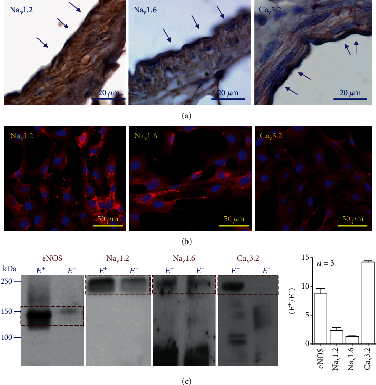
Figure 4.

Expression of voltage-dependent Na+ (Nav) and Ca2+ (Cav) channels in endothelial cells of mesenteric resistance arteries. (a) Immunohistochemistry analysis of the cellular distribution of Nav and Cav channel-specific isoforms Nav1.2, Nav1.6, and Cav3.2 in the wall of mesenteric resistance arteries. Note that Nav1.2 and Nav1.6 channels are present in both endothelial cells and smooth muscle cells, but Cav3.2 channels are expressed exclusively in the endothelium. Arrows highlight the staining observed in endothelial cells. (b) Immunofluorescence detection of the expression of Nav1.2, Nav1.6, and Cav3.2 channels in primary cultures of mesenteric endothelial cells. (c) Representative Western blots and densitometric analysis of the expression of eNOS and Nav1.2, Nav1.6, and Cav3.2 channels in intact mesenteric arteries before (E+) and after (E−) removing the endothelium by the treatment with collagenase.
