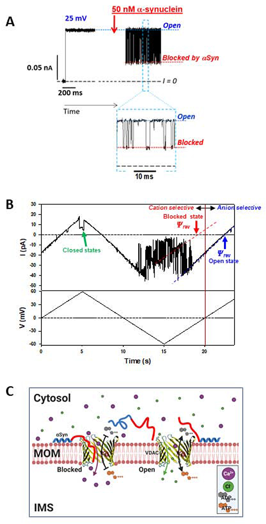
Figure 5. Regulation of VDAC Ca2+ permeability by α-synuclein.

(A) A representative experiment showing a current record through a single VDAC in 1M KCl before and after the addition of 50 nM of α-synuclein (αSyn). αSyn induces fast characteristic blockage events best seen in a finer time scale in Inset. (B) Current through a single VDAC in response to applied triangular voltage wave (50 mHz, ±60 mV) obtained in 150mM/30mM CaCl2 gradient. Dashed blue and red lines indicate VDAC open and αSyn-blocked states, respectively. The intersection of these lines with the zero-current line (I = 0) corresponds to the reversal potential (Ψrev). Positive Ψrev corresponds to anionic selectivity of the open state and negative to cationic selectivity of the αSyn-blocked state. The voltage-induced closed states are indicated by the green arrow. Current records were digitally filtered using a 100 Hz filter. (C) A model of αSyn regulation of Ca2+ flux. Under applied negative voltage, acidic C-terminus of αSyn is captured inside the VDAC pore resulting in reversed VDAC selectivity and consequently in enhanced Ca2+ flux through the pore. When the channel is open, its slight anionic selectivity causes a reduced Ca2+ permeability and favors the fluxes of negatively charged metabolites such as ADP and ATP.
