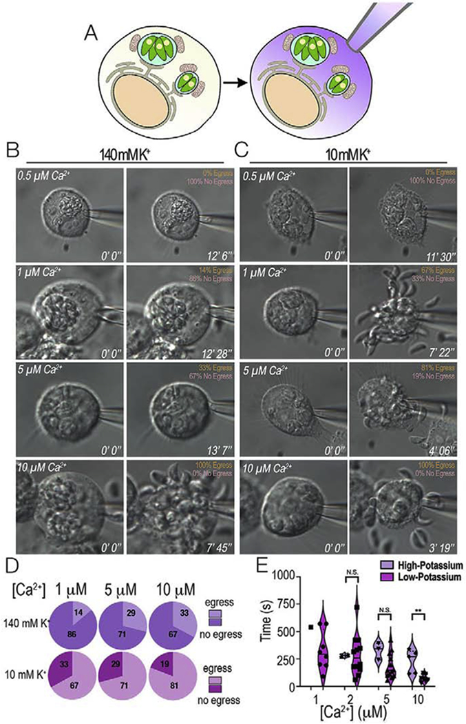
Figure 6: Role of host cytosolic Ca2+ and K+ studied by patching the host plasma membrane.

HeLa cells infected with T. gondii tachyzoites were whole-cell patched and egress was monitored. A, whole-cell patch allowed the exposure of PVs to defined Ca2+ concentrations by exchanging the cytosol of the host cell with the composition of the buffer inside the patch pipette. B, Representative still images of infected host cells patched under high potassium conditions (140 mM K+). Various concentrations of free Ca2+ were tested to monitor egress. The percentage of egressing vs non-egressing parasites is shown in the upper left-hand corner. C, Representative still images of infected hosts cells patched under low potassium conditions (10 mM K+ and 130 mM choline chloride) and egress monitored under the same experimental conditions as in A. D, Percentage of egressing parasites presented as pie charts of increasing Ca2+ concentration. Purple, 140 mM K+, pink, 10 mM K+. E, Violin Plots of the average time to egress under high (140 mM K+) and low potassium conditions (10 mM K+). Note that under low K+ conditions the percentage of egressing parasites increases, and parasites egress faster. N.S. was used to represent non-significant results and ** was used to represent p-values ≤ 0.01 of T-tests between the different Ca2+ concentrations.
