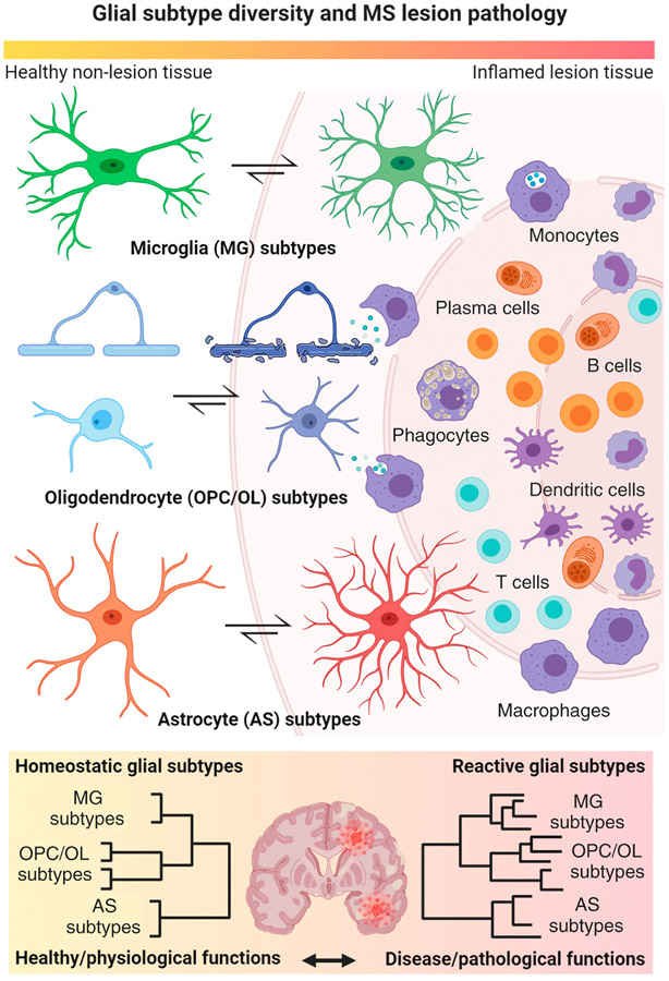
Figure 2. Key Figure. Homeostatic and Reactive Glial Cell Type Diversity in Inflammatory Demyelination.
Main cartoon highlights dynamic transformative states of glial cell types (left center) in response to a focal inflammatory–demyelinating lesion as regularly seen in multiple sclerosis (MS) with blood–brain barrier breakdown and immune cell infiltration into the surrounding parenchyma (right). Note a gradual morphological transformation of glial subtypes from a homeostatic (left) physiological to a reactive pathological state (center) relative to the inflamed lesion area. Under inflammatory–demyelinating conditions, astrocytes, and myeloid cells such as microglia regularly transform into reactive subtypes with enlarged cell bodies and retracted processes. Conversely, oligodendrocyte (OL) lineage cells including OL precursor cells (OPCs) and myelinating cells (OLs) become reactive and exhibit high metabolic stress. Note that most of them undergo cell death close to the demyelinating lesion rim. Lower panel illustrates increased subtype diversity in reactive glial cell populations isolated from inflamed MS brains compared with homeostatic subtype diversity in healthy brains by means of hierarchical clustering with respect to physiological versus pathological subtype functions. This figure was created using BioRender (https://biorender.com/).

