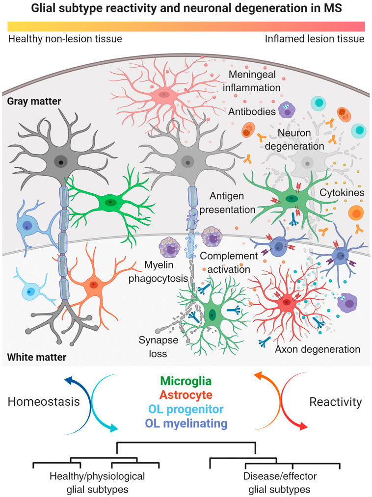
Figure 4. Morphological and Functional Changes in Homeostatic and Reactive Glial Subtypes Relative to Neuron Damage in Multiple Sclerosis (MS) Lesions.
Main cartoon illustrates the sequence of tissue damage in leukocortical (affecting both cortical gray matter and subcortical white matter) MS lesion with presence of infiltrating immune cell types. Note gradual neuronal degeneration from left (healthy state) to right (degenerated state) with axonal degeneration and synapse loss followed by retrograde injury and damage to neuronal cell bodies. Neuron degeneration is paralleled by morphological and functional changes in glial cell types including complement secretion and activation, antigen presentation via MHC class I and II, cytokine production, and complement factor release. Other factors are meningeal inflammation and tissue infiltration of monocyte and lymphocyte subtypes producing antibodies and cytokines, thus further corroborating tissue damage in MS lesion areas. Lower panel demonstrates dynamic cycling of glial subtype states between homeostatic and reactive conditions relative to the inflammatory stage of MS lesions. Note that different glial cells can transform from healthy/physiological subtypes into disease/effector subtypes. This figure was created using BioRender (https://biorender.com/). Abbreviation: OL, oligodendrocyte.

