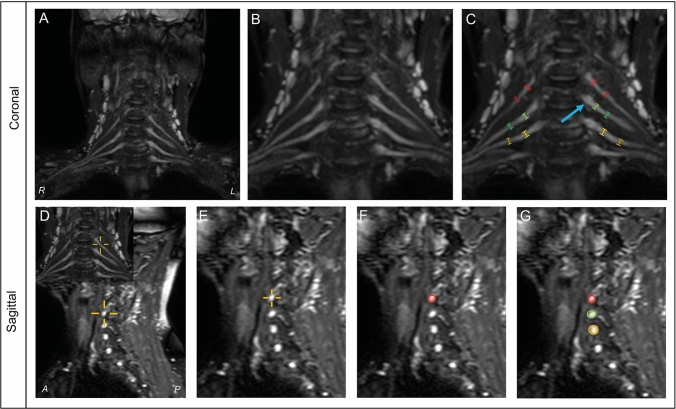
Fig. 1.
Example of nerve root measurements in coronal and sagittal planes. Method of measurements in coronal (upper) and sagittal (lower) planes. Coronal measurements in maximum intensity projection images (a) using 1 × zoom (b) and calipers placed in nerve root C5 (red), C6 (green) and C7 (yellow) next to the ganglion (blue arrow) and 1 cm distal of the ganglion (c). Sagittal measurements in T2 weighted fat-suppressed images using a cross-cursor to identify corresponding measurement sites (d) and 1 × zoom (e). Measurements were then performed at these corresponding measurement sites (f, g). R right; L left; A anterior; P posterior

