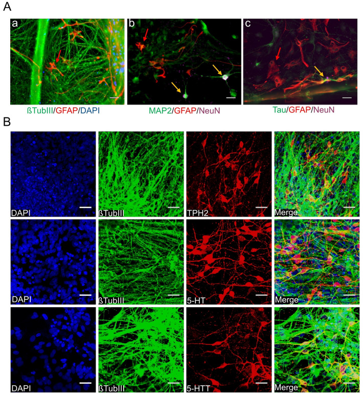
Fig. 2.
Generation of 5-HT specific neurons from human iPSC-derived 5-HT specific progenitors. A Our differentiation protocol generates βTUBIII + neurons (A, a stained in green) and GFAP + astrocytes (A, a–c, stained in red, examples marked by red arrows) following 5 weeks of neuronal maturation. 5-week-old neurons were additionally proven to be mature neurons illustrated by positive staining of (A, b) MAP2 (stained in green) and NeuN (stained in magenta) and (A, c) Tau (stained in green), and NeuN (stained in magenta). Mature neurons are marked by yellow arrows. Pictures were taken using epifluorescence microscopy. B All neurons were co-stained for an antibody against βTUBIII and TPH2, 5-HT, and 5-HTT after 4–5 weeks of neuronal maturation (differentiation week 7–8). Pictures were taken using confocal microscopy. Scale bar: 50 μm. Cell nuclei were counterstained with DAPI and Alexa Fluor 488 (βTUBIII, MAP2, Tau) and Alexa Fluor 555 (GFAP, TPH2, 5-HT, 5-HTT) and Alexa Fluor 647 (NeuN) were used to visualize target proteins. 5-HT specific neuronal differentiation was verified using the JMUi001-A iPS line (data not shown)

