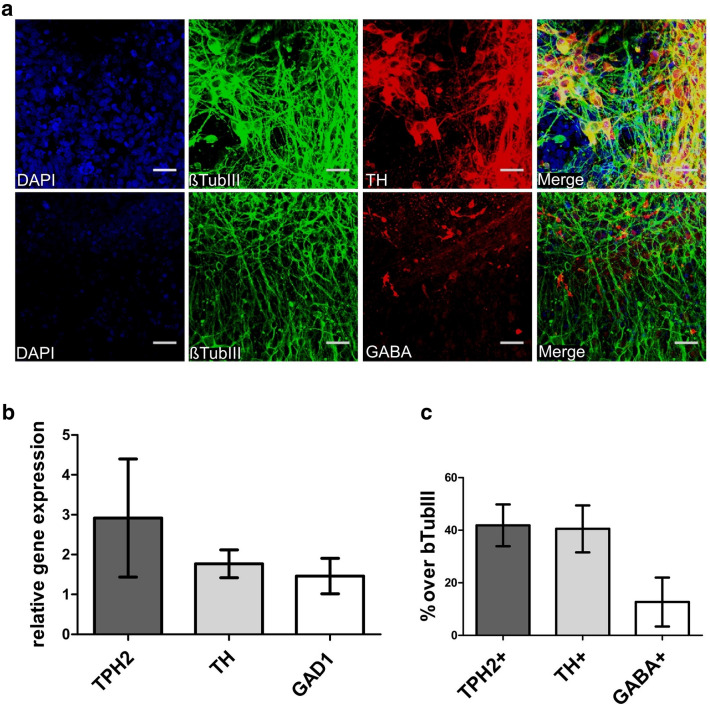
Fig. 3.
Specification of different neuronal subtypes. a Neurons were co-stained for an antibody against βTUBIII, TH, and GABA after 4–5 weeks of neuronal maturation (differentiation week 7–8). Pictures were taken using confocal microscopy. Scale bar: 50 μm. Cell nuclei were counterstained with DAPI and Alexa Fluor 488 (βTUBIII) and Alexa Fluor 555 (TH, GABA) were used to visualize target proteins. b Relative gene expression levels of the human iPSC-derived 5-HT specific (TPH2 +), catecholaminergic (TH +) and GABAergic (GAD1 +) neurons (n = 2 independent differentiations). c Quantification of the amount of 5-HT specific (TPH2 +), catecholaminergic (TH +) and GABAergic (GABA +) neurons within the neuronal cell culture system (n = 3 independent differentiations)

